Laboratory Investigations In Hematological Disoders And Case Study Of Hemoglobinopathies In India

A Medical Laboratory
Clinical Investigations And Red Cell Indices
There are lots of hematological disorders and sometimes differentiating them entails going through lots of laboratory procedures to adequately establish a diagnosis. These procedures aid effectively in ensuring the hematological disease is actually identified and here are the laboratory procedures:
Hemoglobin estimation: It is now accepted that this procedure should be done with internationally accepted standards by the cyanmeth-hemoglobin method using a photoelectric colorimeter. Other visual methods are only approximate at best.
Cell counts: These are made for erythrocytes, leucocytes and platelets. It is also of great significance to knowing the differential counts of leucocytes.
Reticulocyte count: It is done directly in the wet preparation after staining with brilliant cresyl blue which is a vital stain.
Estimation of packed cell volume (PCV)-venous hematocrit.
The red cell indices: These are calculated from packed cell volume (PCV), hemoglobin (Hb) and red cell count. They are MCHC, MCH and MCV.
- MCHC: Mean corpuscular hemoglobin concentration represents the concentration of hemoglobin in the red cell. It is calculated as follows: MCHC = (Hb in g/dL X 100)/PCV (g/dL). It’s normal range is 32 to 36 g/dL.
- MCH: Mean corpuscular hemoglobin represents the average weight of hemoglobin in each cell and is calculated as follows: MCH = (Hb in g/dL X 10)/RBC count in million/cmm (pg). It’s normal range is from 27 to 32 pg (pictogram).
- MCV: Mean corpuscular volume gives the average volume of red cells and is calculated as follows: MCV = PCV X 10/RBC count in million/cmm (fl). It’s normal value varies from 76- 94 fl (femtoliters).
Hematological Investigations
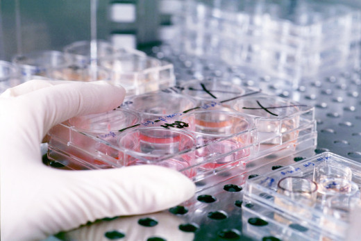

Special Hematological Investigations
Osmotic fragility of erythrocytes: It is determined by estimating the hemolysis when a standard volume of erythrocytes is added to increasing dilutions of sodium chloride. Osmotic fragility is increased in spherocytosis.
Peripheral blood smear: This is the most useful single investigation which will help in diagnosis. All the cellular elements are examined systematically. Erythrocytes may be small (microcytes), large (macrocytes) or normal in size (normocytes). Variation of size is called anisocytosis. Poikilocytosis refers to difference in shape. The cells may be pear-shaped, elliptical, or sickle-shaped. Schistocytes are fragmented red cells. Acanthocytes (spur cells) show numerous spiny projections. Both are seen in hemolytic anaemias. Burr cells show short projections on their surface. These are seen in renal failure.
Bone Marrow biopsy: This is an important and integral part of hematological examinations. Bone marrow examination is absolutely necessary to establish the diagnosis of leukemias, myeloma, myelofibrosis, hypersplenism, lymphomas, aplastic anaemia, megaloblastic anaemia and other dyshemopoietic states. It is helpful in the diagnosis of kala-azar, histiocytosis, amyloidosis, secondary carcinomatosis and several other conditions.
The sites for bone marrow aspiration include the sternum, posterior and anterior portions of the iliac crest, vertebral spines, ribs and shaft of tibia. The conventional Cox needle or the Jamshidi needle are used at present. Injury to mediastinal structures may result if sterna puncture is done without proper care.
Hemoglobinopathies

A General Overview
Hemoglobinopathies are among the most widespread genetic disorders seen in humans. The hemoglobin in normal individuals is made up of three components. These are Hb A (94-97%); Hb A2 (1-3.5%) and Hb F (0-2%). During fetal life, Hb F is the main hemoglobin and it is replace by Hb A within the first six months of life. Failure of the formation of adult hemoglobin to replace the fetal hemoglobin results in thalassemias. This transformation is under genetic control.
The hemoglobin molecule is made up of heme and globin. Chemical changes in the heme lead to the production of methemoglobin, suphemoglobin and carboxyhemoglobin. The globin is made up of two types of polypeptide chains: Hb A has 2 alpha and 2 beta, Hb F has 2 alpha and 2 gamma, and A2 has 2 alpha and 2 delta chains. Each polypeptide chain has a specific amino acid constitution. When the sequential order of amino acids is latered or the chain is altered by deletion or addition, abnormal hemoglobin are formed. For example, substitution of valine for glutamic acid at sixth residue of beta chain results in the formation of Hb S and lysine for glutamic acid results in the formation of Hb C. Such alterations result in the change of electrical charge of the molecule and consequently, the electrophoretic mobility alters. This property enables these abnormal hemoglobins to be separated by electrophoresis. Abnormal hemoglobins and thalassemia are inherited as autosomal dominants, but their expressivity and penetrance vary considerably. Several variants of hemoglobin A have been identified.
Several Variants Of Hemoglobin In India

Hemoglobinopathy in India
Several hemoglobin variants are encountered in India. These are hemoglobins D, E, F, H, I , J, K, L, M, S, Q, Norfolk and Lepore. Of these, prevalence of only hemoglobins S, E, D, J, K and Q have been studied in detail in population groups. Hemoglobin S is found mostly in the tribals from Tamil-Nadu, Madhya Pradesh, Bihar and Orissa; Hb E is seen in Bengal and Assam; Hb D, J and K are seen in Punjabs and Gujaratis and Hb Q Is seen in Sindhis. Among these, only Hb S and Hb E are widely prevalent. Hemoglobin C is not reported from India.
Hemoglobinopathies lead to physical and chemical alterations in the function of the hemoglobin molecule depending on the site and nature of the amino acid substitution. Many lead to hemolytic anemia, eg, Hb S. Substitution of certain nonpolar amino acids within the hemoglobin molecule makes it unstable so that even in the heterozygous state it leads to hemolytic anemia. Certain substitutions lead to congenital methemoglobinemia.
Some substitutions lead to increased affinity of the hemoglobin to oxygen to the tissues. Tissue anoxia leads to polycythemia. Abnormal hemoglobins can be separated by electrophoresis in suitable buffers. In comparison with the mobility of hemoglobin A, they can be classified as slow moving, fast moving and very fast moving types.
India's epidemiology is an excellent case study since its occurrence is overwhelming and gross when compared to other parts of the world.
© 2014 Funom Theophilus Makama


