Oesophageal Diverticulum Health Implication: The Story Of Thomas Putressi From Christmas Island

Oesophageal Diverticulum
The Pouch in Oesophageal Diverticulum

Thomas Putressi's Story
NOTE: Additional Information in the coloured columns are more medically inclined for medical personnels and hence not really necessary for all to read.
Thomas Putressi lives in Poon Saan in Christmas Island. He is 54 years old and often suffers from Inflammation of the Oesophagus. For the past 20 years, he has been experiencing pain around his Oesophagus and difficulty in swallowing at least once every year. In the first instance he was diagnosed with Oesophagitis, which is the inflammation of the Oesophagus and that is why he suffered from such symptoms, earlier listed. But surprisingly, for the past 2 years, he did not experience any, not until some weeks back. But this time it manifested quite differently.
Mr Thomas started experiencing the usual pain on his Neck region which gradually became intense. Three days later, he experienced difficulty in swallowing food and then cough. It did not stop there, the following day, he had serious bad breath and when he woke up in the morning, he noticed his pillows were wet, which became a trend in subsequent nights. But also during the day time, he salivates a lot. After about a week of such symptoms, he went to the Hospital, was given a substance called Barium with a meal, after which an X-ray examination was taken. Not only that, a long thin tube was passed through his gastro-intestinal tract for some diagnostic examination, after which the Doctor's diagnosis stated:
True, Acquired Multiple Oesophageal diverticula, Uncomplicated form at the level of Bifurcation of the Trachea
Hell No! what is this?
Anatomy of the Esophagus

Histology of the Oesophagus
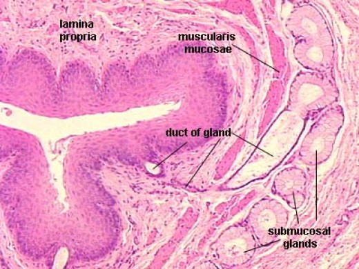
The Oesophagus has 4 layers
- The Mucosa layer:It is a non-keratinized stratified squamous epithelium: It is rapidly turned over and serves as protection due to high volume transit of food, saliva and mucus. The Mucosa layer consists of two sub-layers, which are: Lamina Propria :Sparse- constituent of the most linings known as mucous membranes or mucosa; it is a thin layer of loose connective tissue which lies beneath the epithelium and together with the epithelium constitutes the mucosa. And the,Muscularis Mucosa: Contains the mucous secreting glands (Oesophageal glands) and connective structures.
- Submucosa: Containes the Mucous secreting glands (Oesophageal glands) and connective structures called Papillae.
- Muscularis externa: Composition varies in different parts of the Oesophagus, to correspond with the conscious control over swallowing in the upper portions and the autonomic control in the lower portions. Upper one-third or the superior part consists of the straited muscles: middle one-third consists of smooth muscles and straited muscles (mixed) and finally, the inferior one-third is predominantly smooth muscles.
- Adventitia: Outermost connective tissue covering of any organ, vessel or structure.
These are the histological layers of the Oesophagus.
Histological layers of the Oesophagus
What is Oesophageal Diverticulum
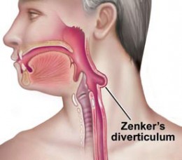
If we look at the first picture, we can see a diagram of an individual with an Oesophagus which has a "baggy" extension or a pouch. So, Oesophageal Diverticulum is the outpouching of the Oesophageal wall, filled with mucus and undigested food.
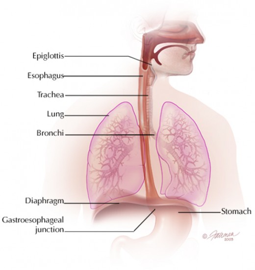
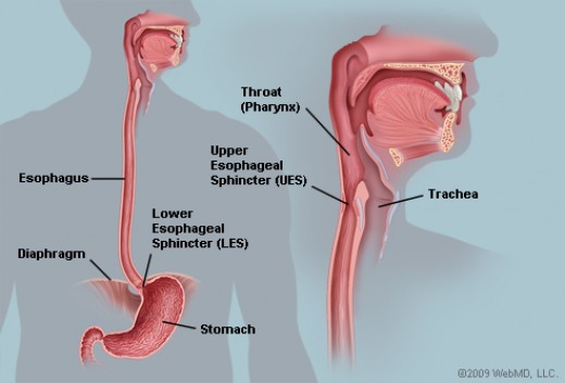
Now, look at the second diagram very closely. You can see the Oesophagus which runs through from the Mouth to the Stomach like a tube. There is the region where it pierces the Diaphragm to enter the stomach, in this region is a sphincter Muscle (just at the Oesophageal-Stomach junction). As we already know, the essence of the Oesophagus is for the transportation of food from mouth to the stomach for digestion and this transport ought to be one way (from mouth to Stomach). The movement of the Oesophageal muscles which enables this to occur is known as Peristalsis and the Sphincter muscle at the Oesophageal-Stomach junction ensures that food does not go back to the Oesophagus when already in the stomach. If we fully understand this, we can then proceed.
This sphincter muscle helps maintain a certain pressure in the Oesophagus called the intra-Oesophageal pressure. Sometimes, this pressure increases and when it does, it exerts itself on the walls of the Oesophagus making these walls expand continuously and resulting to their weakness. If this continues, the region(s) of weakness of the Oesophageal wall begin to herniate and as this continues an extra extension in form of a pouch (as in first picture) is formed. By the time this pouch begins to house undigested food and accumulate mucus, Oesophageal diverticulum is formed.
The increase in the intra-Oesophageal pressure could be due to prolonged irritation of the Oesophageal walls as in Oesophagitis (in the case of Mr Thomas Putressi) or Inflammatory processes in nearby organs of the Oesophagus (para-Oesophageal inflammation) or processes which lead to lack of blood and nutrient supply to the Oesophagus (Sclerotic processes). The last situation leads to the traction of the Oesophagus to other nearby Organs (more often the right bronchus, which is the closest to the Oesophagus as we can see in picture 2) thereby forming Oesophageal pouches. In this case it is calledTraction diverticula. Or it could also be due to regular pressure exerted by solid food passing through to the stomach. In this case, it is called Pulsion diverticula.
Anatomical Classifications of Oesophageal Diverticulum
What Are the Forms or Classifications of Oesophageal Diverticulum?
Let us focus on Photos 4 and 5 very closely [You can click on the photo for a clearly or larger view]. In 5 especially, it is clearly indicated that the Pharynx which is a tube from the mouth extends down inside the body and continues as the Oesophagus (anatomical location is C6). The Junction where the Pharynx becomes the Oesophagus is known as the Pharyngo-Oesophageal junction and this is a very common place where Diverticulum occurs. In fact, this type of diverticulum is what is commonly called the Zenker's diverticulum. Let's focus more on Photo 4 now, we can notice where the trachea bifurcates (or divides) into left and right bronchi. The area of this junction of bifurcation on the Oesophagus is another point where diverticulum occurs. This location is also exactly at the middle of the chest or thoracic region, so it is also called the Mid-chest or Mid-thoracic region. The final Anatomical location of pouch formation is where the Oesophagus pierces into the Diaphragm as it enters the stomach (first picture and Photo 4). On the Oesophagus, just above the piercing is this common site for pouch formation of Oesophageal diverticulum and this is known as Epiphrenic diverticula (above the diaphragm).
From the additional information on the blue column, we can see that the Oesophagus when observed or studied histologically (microscopically) consists of 4 layers. When two layers only are affected, it is called a False diverticulum, but if the entire 4 layers are affected, then it is True diverticulum (One major false diverticulum is Pseudodiverticula which occurs due to herniation of mucosa and submucosa layers through a defect of the muscular wall, for example the Zenker diverticulum).
Also, this ailment could occur right from birth. In this case it is known as congenital Oesophageal diverticula. But if suffered during Life (not from birth), then obviously, it is an acquired disease. More forms of this ailment is: the pouch could be just one (single) or more than one (multiple). Finally, it could come with complications, for example, bleeding or it can also be without such complications. Usually, pouches less than 2 cm in size rarely lead to complications.
Now that we know the various classifications or forms, we can carefully analyze the diagnosis of Mr Thomas Putressi. It is a true form because all histological layers of the Oesophagus are involved; acquired because he did not suffer this ailment from birth, multiple because the pouch isn't just one, bifurcation of the Trachea is the location of the numerous pouches and the diverticulum is without complication and hence the diagnosis: True, Acquired, Multiple Oesophageal diverticula. Location of area of Bifurcation and uncomplicated form.
We can quickly review the classification again:
- according to affection of layers: True and false.
- according to occurrence: Congenital and acquired
- according to number of pouches: Single and multiple
- according to location: Pharyngo-esophageal (Zenker's), bifurcational (mid-thoracic or mid-chest) and Epiphrenic diverticula (above the diaphragm).
- According to clinical course: complicated and uncomplicated.
Oesophageal Diverticula

Treatment
Treatment is only surgery. Though in the case of bifurcational diverticula (mid-thoracic or mid-chest), operation is indicated only in one out of ten patients. In such conditions, Oesophageal diverticula must have been complicated with bleeding, perforation, Oesophago-Bronchial fistula or even malignancy. So in such a case, conservative therapy is needed; especially strict lifestyle change such as: eating balanced diet, chewing food thoroughly and drinking of plenty of water after meals to wash off any form of food remnant. if symptoms still persist, surgery becomes the only option.
Treatment of diverticula requires:
- Thorough evaluation of diverticula
- repair of the weakened wall;
- relief of obstructions.
Stages of Operation
- Mobilization of the diverticulum
- suturing of its base
- suturing of the muscular sheath over the surface line
Surgeries
- Endoscopic diverticulotomy (Dohlman procedure): This procedure divides the septum between the cervical Oesophagus and diverticular Pouch. By doing so, food can drain freely to the Oesophagus from the pouch.
- Cricopharyngeal Myotomy: Removal of small diverticula; such operation occurs using open or trans oral approach.
- Diverticulopexy with cricopharyngeal myotomy: Removal of larger diverticular; involves turning the diverticular sac upside down and suturing it to the Oesophageal wall to suspend it.
- Diverticulectomy and Cricopharyngeal myotomy: For the treatment of Zenker's diverticula involves a complete excision of the diverticular sac, indicated for diverticula greater than 4 cm in size.
Accesses
In order to expose Pharyngo-Oesophageal diverticula; the cervical access along the anterior border of the sternocleidomastoid muscle is applied; in case of bifurcational diverticula, right-side posterio-lateral thoracotomy in IV intercostal space is performed in epiphrenal diverticular left side posterolateral thoracotomy in VII intercostal space.
Signs, Symptoms, Complications And Risk Factors
1. Hyper-salivation: At night, the patient wets his/her pillow, just as in Mr Thomas case. A sign clinically known as "Wet Pillow" symptom which is due to increased salivation and nocturnal effluence of saliva and mucus from the mouth.
2. Dysphagia: Difficulty in swallowing is known as dysphagia. This is also characterized by a feeling of food caught in the throat. Sometimes a compressible mass can be found on the Neck, usually on the left side. The patient will need to press this mass to swallow food or make unusual neck movements in order to empty the diverticulum.
3. Bad breath and gurgling sound: Since a pouch is formed in this pathology, food sometimes can enter the pouch and remain there for long period of time, resulting to decay and smell. This smell is what accounts for the bad breath as in the case of Mr Thomas. The gurgling sound happens when the patient eats. This gurgling sound is called Boyce's sign.
4. Cough: This occurs usually in advance stages of the disease.
5. Sometimes Neck pain and weight loss.
Oesophageal diverticulum can come with complications. The pouch formed can house food materials which can later decay, irritate it and cause inflammation known asdiverticulitis.
Also, the wall of the Oesophagus (due to the increase in intra-oesophageal pressure) can tract with the Trachea, both walls joining together and forming an opening. This case is called Oesophageal-Tracheal fistula. It doesn't just end there, when food is consumed, it can pass through the opening formed and hence go into the Trachea. Since the Trachea is meant for breathing (passing of air), food entering into it becomes a pathology called Pulmonary aspiration which is dangerous (especially in children). Oesophageal-Tracheal fistula can also cause infection of the lungs (Aspiration Pneumonia).
Also, bleeding can be another complication of this pathology.
Risk factors
Risk factors are those situations which make individuals vulnerable or susceptible to suffering from a disease. The following factors put individuals more at risk in getting or acquiring Oesophageal diverticulum:
- Individuals who are 50 years and above. Mr Thomas Putressi is 54 years old, and so is well defined by this factor.
- People suffering from chronic inflammation of the Oesophagus (Esophagitis) are at a very high risk (also as is the case with Mr Thomas Putressi).
- Patients suffering from Oesophageal motility (movement) disorders are at a high risk of suffering from Epiphrenic Diverticula (above the Diaphragm) because the lower Oesophageal Sphincter in the border between the stomach and the Oesophagus (Muscles of the contractile ring) fail to relax normally during swallowing.
- Gastro-Oesophageal reflux (reflux of acid from the stomach into the Oesophagus) is another risk factor especially for Zenkers and mid-thoracic diverticula.
- Hiatal Hernia (bulging of the stomach past the diaphragm into the chest cavity) is another risk factor, especially for Zenkers diverticula.
Zenker's diverticulum
. The pouch is a single one.")

Diagnosis and Differential Diagnosis
- Barium Swallow: Barium preparation in the form of liquid or other forms is swallowed and its movement through the Oesophagus is analysed via X-ray technology.
- Gastrointestinal tract Endoscopy or Fibrogastroduodenoscopy: The usage of a narrow and flexible tube called an endoscope which is passed through the G.IT and projects images of the internal structures of the Oesophagus and projects them into a screen. This was used in Mr Putressi's case.
- Oesophageal manometry: Tests and measures the Oesophageal strength and timing of contractions and relaxation of muscular valves.
- 24-hour pH metry: To exclude any suspicion on gastro-Oesophageal reflux diseases (GERD).
Differential diagnosis
- Pseudodiverticula (Functional Diverticula): In this case, dysphagia will be intermittent which will usually arise only after meals or strong excitements.
- Sternocardia: Retrosternal pain attacks with irradiation in the left arm and left shoulder blade (left scapula) is peculiar to it. Usually, the pain disappears after Nitroglycerin administration. Another important clinical issue is the feeling of fear. But in Oesophageal diverticulum, there is no obvious sensation of fear, and the retrosternal pain caused by the spastic stricture or diverticulum of Oesophagus is characterized by feeling of compression deeply inside which is accompanied by disturbances of swallowing, sometimes vomiting after which pain disappears.
This was exactly how Mr Thomas Putressi was diagnosed and treated. You can find the treatment plan in the second coloured column of this hub. Endeavour to leave your questions and comments. I hope I was able to explain this ailment as simple as possible for all to comprehend.
© 2014 Funom Theophilus Makama
Related

Acid Reflux Symptoms and Natural Remedies

Symptoms of Appendicitis, and Ruptured Appendix....know what to look for.

Delayed Gastric Emptying in Children: Our Family's Experience

Mastocytic Enterocolitis: Symptoms, Causes, Complications, Diagnosis, Treatment, Prevention

Miracle Man : My Son Has Wegener's Granulomatosis