Echinococcosis: General Health Implications, Morphology, Lifecycle And Clinical Manifestations

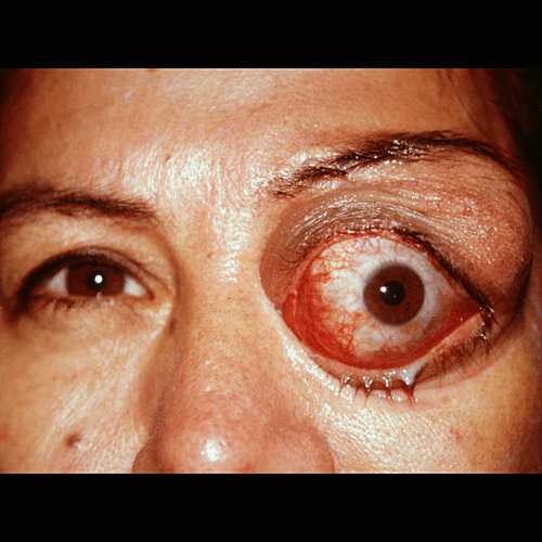
Echinococcosis Manifestation On The Eye
A General clinical Overview Of Echinococcosis
Infection of human tissues by larval form of Echinococcus granulosus and less commonly E. multilocularis is termed echinococciasis. The disease is worldwide in distribution. It is more common in sheep-rearing countries both in the tropics and temperate zones. Close contact between man, dog and sheep favours this infection. Echinococcus granulosus is widely distributed in India and the disease is seen endemically or sporadically.
Morphology: The adult worm is small, measuring only 3 to 6 mm in length. The scolex bears four suckers and rostellum with two rows of hooklets. There are only three segments. The last segment is mature and gravid. Dogs pass the ova in their feces. The ova resemble those of t. saginata and contain the infective embryo.
The larval form is the hydatid cyst seen in tissues. The hydatid cyst is formed when the larva reaches the tissues in the blood stream and develop further.
Life cycle: Definitive hosts are the dogs, wolves, jackals and other canines. Intermediate hosts are sheep, pigs, goats and humans. Sheep are the optimum intermediate hosts and they serve to perpetuate the natural lifecycle of the parasite.
The eggs passed in dog feces are ingested by the intermediate hosts. The embryos are liberated in the intestines and they penetrate the mucosa and enter the portal radicles to reach the liver. The larvae are arrested mainly in the liver and lungs but some may escape into the systemic circulation to reach other viscera like the brain, bones, kidneys, etc. 5 to 10 mm in diameter and these are known as hydatid cysts (hydatids= drops of water). They grow to reach large sizes and the parasite multiplies in number.
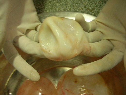
The hydatid cysts has a thick fibrous outer wall and an internal germinal layers, from which brood capsules containing budding scolices arise. These may separate from the capsule and float inside as daughter cysts. The hydatid fluid is a clear colourless fluid which is highly antigenic. On keeping it a granular deposit forms at the bottom (hydatid sand) which contains free scolices and brood capsules.
When the hydatid cysts or scolices are ingested by the definitive hosts, they attach to the intestinal mucosa and grow into adults. Each animal may harbour many worms.
Removal Of Cyst Membrane In The Treatment Of Echinococcosis
Infectious Diseases

Clinical Manifestations Of Echinococcosis
Infected dogs are asymptomatic. In human hydatid disease, symptoms are due to allergic reactions and mechanical compression of the surrounding tissues. Local symptoms depend on the site and size of the cyst. Clinical features are those of an enlarging mass. Commonest presentation is with hepatomegaly, hemoptysis or neurological symptoms. In about 75% of cases, the right lobe of the liver is the seat of large cysts. Lungs, brain, orbits and bones are affected not uncommonly. An interesting physical finding is the hydatid thrill which is elicitable by percussion over large hydatid cysts. The presence of free brood capsules in the fluid gives the sensation on the pleximeter finger, which confirms the diagnosis.
Allergic manifestations occur either as nonspecific allergu such as urticaria, eosinophilia and rashes. Sometimes, rupture of a cyst and entry of the fluid into tissues evoke anaphylactic reactions.
© 2014 Funom Theophilus Makama


