Different Radiographic Representation Of Hemothorax: It's Manifestation As Seen On The X-ray

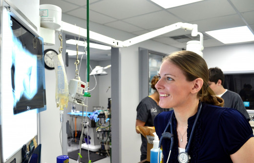
X-Ray Diagnostics
Introduction
Hemothorax is as a result of Trauma to the chest cavity or thoracic region. When this occurs, the pleural space may experience some tear and hence the presence of blood in it. The source of blood could be from any part of the thoracic region be it vessels, the heart, chest wall or the lung parenchyma itself.
Treatment is only done through surgery and this procedure is called, tube thoracostomy. A tube is inserted into the pleural to remove the blood accumulated in it. When inserted, the lungs expand and the bleeding stops. Sometimes, the blood accumulated may clot and result to what is known as retained hemothorax. In such a situation, highly functional tubes are used to remove the blood.
This hub serves to show pictorial representations (On X-ray) only as there are already lots of articles around the web which talk about hemothorax. This situation is an emergency one which can be treated only via surgery.
Hemothorax On X-ray


 may be used. Thoracotomy is the procedure of choice for surgical exploration of the chest when massive hemothorax or persistent bleeding is prese")


 can be responsible. Blunt or penetrating injury involving virtually any intrathoracic structure can result in hemothorax.")


References
1. All X-ray photos here are from DFM E-Group, in the photo and X-ray section.
2. Notes and explanations: Wikipedia, Essentials of Clinical Medicine by Kumar and Clark's, Medicinenet and Mayor's Clinic.
© 2014 Funom Theophilus Makama

: Identifying A Normal Chest X-Ray")

%20may%20be%20used.%20Thoracotomy%20is%20the%20procedure%20of%20choice%20for%20surgical%20exploration%20of%20the%20chest%20when%20massive%20hemothorax%20or%20persistent%20bleeding%20is%20prese)
%20may%20be%20used.%20Thoracotomy%20is%20the%20procedure%20of%20choice%20for%20surgical%20exploration%20of%20the%20chest%20when%20massive%20hemothorax%20or%20persistent%20bleeding%20is%20prese&picture=https://usercontent1.hubstatic.com/8717586.jpg&redirect_uri=https://hubpages.com/health/Different-Radiographic-Representation-Of-Hemothorax-Its-Manifestation-As-Seen-On-The-X-ray)
%20can%20be%20responsible.%20Blunt%20or%20penetrating%20injury%20involving%20virtually%20any%20intrathoracic%20structure%20can%20result%20in%20hemothorax.)
%20can%20be%20responsible.%20Blunt%20or%20penetrating%20injury%20involving%20virtually%20any%20intrathoracic%20structure%20can%20result%20in%20hemothorax.&picture=https://usercontent2.hubstatic.com/8717589_f520.jpg&redirect_uri=https://hubpages.com/health/Different-Radiographic-Representation-Of-Hemothorax-Its-Manifestation-As-Seen-On-The-X-ray)