Clinical Significance Of Blood Formation In Basophils, Monocytes, Thrombocytes And Lymphocytes

A Basophil
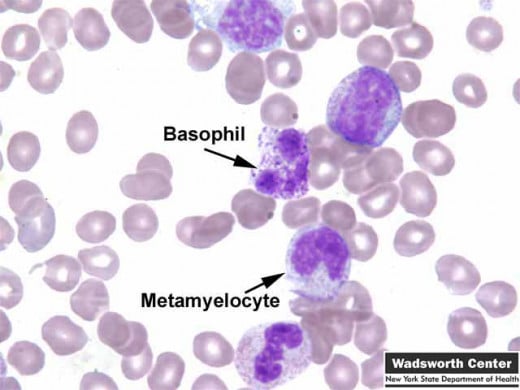
Basophils
Introduction
Leucocytes are white blood cells responsible for protective actions against foreign bodies infiltrating the human body. They are the Neutrophils, Basophiles, Eosinophils, monocytes and lymphocytes. Already discussed in this series are the Neutrophils and Eosinophils, hence we will continue with the other leucocyte cell types. Another type of blood cells are the Plasma cells (Thrombocytes) which are largely responsible for clotting of blood to prevent excessive bleeding.
BASOPHILS
Basophils form up to 1% of the total leucocytes. They are identified by the presence of coarse basophilic granules which often overlie the nucleus. The granules are rich in heparin and histamine. In the tissues they develop into mast cells. They possess sites of attachment of IgE and they release histamine on degranulation. Increase in basophil leucocytes (counts above 0.1 X 10^9/liters) is uncommon, but this is seen in myeloproliferative disorders such as chronic myeloid leukemia and polycythemia vera. Disorders like myxedema, smallpox, chicken pox and ulcerative colitis also may be associated with basophil leucocytosis.
Lymphocytes View From Electron Microscope
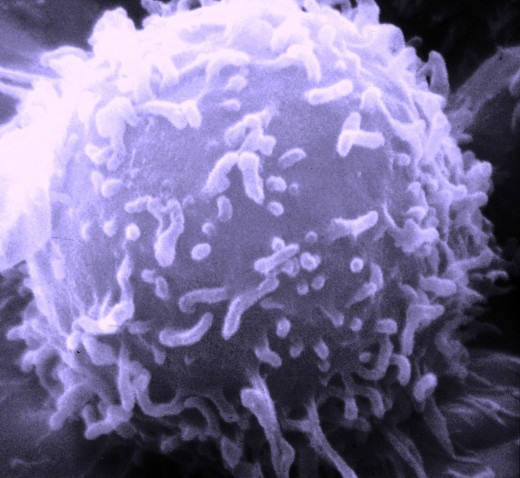

Lymphocytes
Lymphocytes and plasma cells are together known as immunocytes. Their role is to assist the phagocytes in defence mechanism and add specificity to the attack. Lymphocytes are formed in the bone marrow and thymus during postnatal life. In the fetus, the yolk sac and liver produce lymphocytes. In the lymphopoetic tissues, the stem cells undergo spontaneous divisions without depending on antigenic stimulation. Other lymphoid tissues found in lymph nodes, speen, organized lymphoid tissues of the alimentary and respiratory tracts and the lymphocytes seen in blood and tissue spaces constitute secondary lymphoid tissues.
Most of the lymphocytes in peripheral blood are small with scanty nongranular cytoplasm and a central nucleus with coarse chromatin. Larger lymphocytes which may occur in peripheral blood are probably stimulated by antigenic challenge from viruses or foreign proteins.
B lymphocytes are derived from bone marrow stem cells. When activated by antigens, the B cells proliferate and mature into plasma cells which secrete specific immunoglobulins, the antibodies. The primordial T cells are probably produced in the bone marrow and possibly the cortex of the thymus, where they are conditioned to recognize the body’s antigenic make-up. They are the main cells responsible for cell-mediated immunity. Helper T cells are specific lymphocytes which instruct B cells about specific antigens. The T cell population also contains suppressor cells which reduce B cell responses. The killer cells (K cells) cause damage to cells recognized as ‘foreign’. Lymphocytes migrate to the lymph nodes and spleen through post-capillary venules. The T cells are seen in the perifollicular areas of the cortex of lymph nodes (para-cortical areas) and in the periarteriolar sheaths surrounding the central arterioles of the spleen. The majority of lymphocytes in the thoracic duct and peripheral blood are T cells. B cells selectively accumulate in the subcapsular periphery of the cortex and medullary cords of lymph nodes and germinal follicles of lymph nodes and spleen. Majority of B cells remain for long periods in these sites.
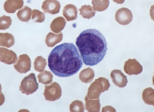
Monocytes
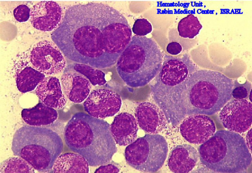
Plasma Cells
Monocytes And Plasma Cells
MONOCYTES
Monocytes arise from stem cell precursors present in the marrow, through the stages of monoblasts and promonocytes. From the marrow, they are released and after circulating for 20-40 hours, in the peripheral blood, they enter the tissues where they mature. Their principal function is phagocytosis and they function as tissue macrophages. In the tissues, their lifespan varies from several months to even years. Bacterial infections such as tuberculosis, brucellosis, bacterial endocarditis, typhoid and protozoal infections like amoebiasis may lead to moderate monocytosis (over 0.8 X 10^9/liter). Other causes of monocytosis are neutropenia, Hodgkin’s disease, and monocytic leukemia.
PLASMA CELL SERIES
Plasma cells form about 2-3% of cells in the bone marrow. Though they are not usually seen in the peripheral blood, they may appear occasionally. Plasma cells accumulate in chronically inflamed tissues. Such plasma cells contain eosinophilic (acidophilic) smooth hyaline spherical cytoplasmic bodies called Russell bodies. These bodies are composed mainly of gamma-globulin. Plasma cells constitute the main source of antibodies. They are formed from stem cells in the bone marrow and then migrate to other tissues. Plasma cells synthesizing IgA migrate to gastro-intestinal tissues while cells producing IgG and IgD go to tonsillar tissue.
© 2014 Funom Theophilus Makama
Related

Donating Blood--History, Statistics, Facts and More

Facts about Blood/Blood Cells Facts: Function of whole Blood, function platelets, function red/white blood cells

Nano Robots - Tiny Futuristic Tools for Medical Science

The Importance of Water in the Human Body

The Thymus Gland, T Cells, AIDS, and Myasthenia Gravis


